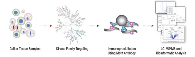
The reversible addition of phosphate groups by protein kinases can activate or inhibit protein function. Phosphorylation signaling cascades, for example the MAP kinase signaling pathway, are important for transmitting information from outside to inside the cell where it ultimately results in modulation of gene transcription. Aberrant phosphorylation is widespread in diseases including cancer, diabetes, and neurodegeneration. Therefore, identifying the phosphorylation status of proteins is crucial to understanding cellular processes in both health and disease.
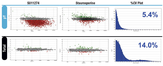
Profiling tyrosine phosphorylation in MKN-45 cells. Peptides from MKN-45 cells treated for 2hr with DMSO (control), 1 μM SU11274 ,or 200 nM Staurosporine #9953 for were enriched using the PTMScan® Phospho-Tyrosine Rabbit mAb (P-Tyr-1000) Kit #8803 (above). In parallel, total protein levels were profiled by running unenriched material in LC-MS/MS (below). Peptides that decreased in abundance with SU11274 or Staurosporine are indicated in red, peptides that increased with treatment in green. A % CV histogram of analytical replicates for each enrichment is shown on the right with the median % CV indicated in blue.
Note: Custom mixes of Ser/Thr motif antibodies can be made for services. Please inquire.
| Target Description | Motif | Reference Data |
|---|---|---|
| Target Description | Motif | Reference Data |
| 14-3-3 Binding Motif | (R/K)XX(s/t)XP | |
| Akt Substrate | RXX(s/t) | Mouse Liver | XLS | RAW |
| Akt Substrate | RXRXX(s/t) | Mouse Liver | XLS | RAW |
| AMPK Substrate | LXRXX(s/t) | Mouse Liver | XLS | RAW |
| ATM/ATR Substrate | (s/t)Q | Mouse Liver | XLS | RAW |
| ATM/ATR Substrate | (s/t)QG | Mouse Liver | XLS | RAW |
| CDK Substrate | (K/R)(s/t)PX(K/R) | |
| CK2 Substrate | (s/t)(D/E)X(D/E) | |
| MAPK/CDK Substrate | PX(s/t)P, (s/t)PX(K/R) | Mouse Liver | XLS | RAW |
| PDK1 Docking Motif | (F/K)XX(F/Y)(s/t)F/Y) | Mouse Liver | XLS | RAW |
| PKA Substrate | (K/R)(K/R)X(s/t) | Mouse Liver | XLS | RAW |
| PKC Substrate | (K/R)X(s/t)X(K/R) | Mouse Liver | XLS | RAW |
| PKD Substrate | LXRXX(s/t) | |
| PLK Binding Motif | S(s/t)P | Mouse Liver | XLS | RAW |
| tP Motif | (s/t)P, (s/t)PP | |
| tPE Motif | (s/t)PE | Mouse Liver | XLS | RAW |
| tXR/tPR Motif | (s/t)XR, (S/t)PR | Mouse Liver | XLS | RAW |
| Phospho-Tyrosine (pY-1000) | y | Mouse Brain | XLS | RAW |