| Cat. # | Size | Qty. | Price |
|---|---|---|---|
| 63387T | 1 Kit (9 x 20 microliters) |
|
| Product Includes | Quantity | Applications | Reactivity | MW(kDa) | Isotype |
|---|---|---|---|---|---|
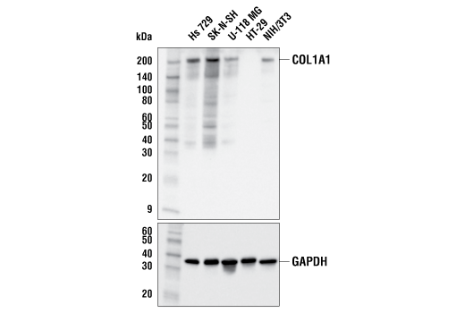
| COL1A1 (E8F4L) XP® Rabbit mAb 72026 | 20 µl |
|
H M | 220 | Rabbit IgG |
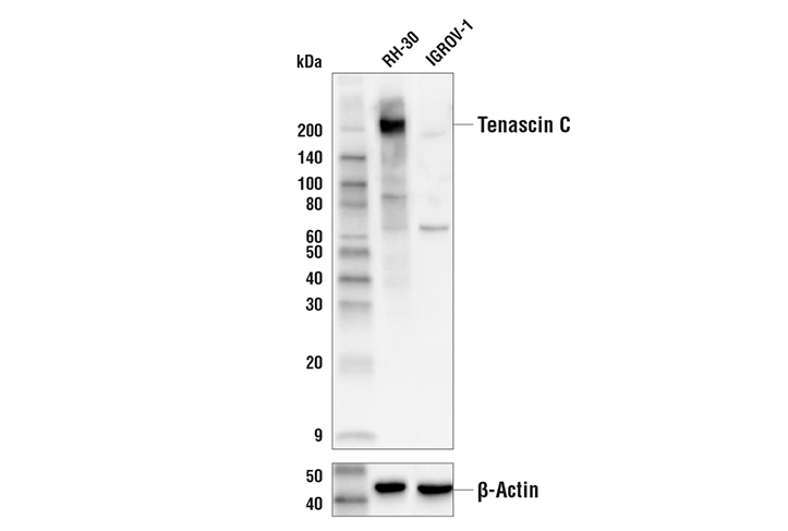
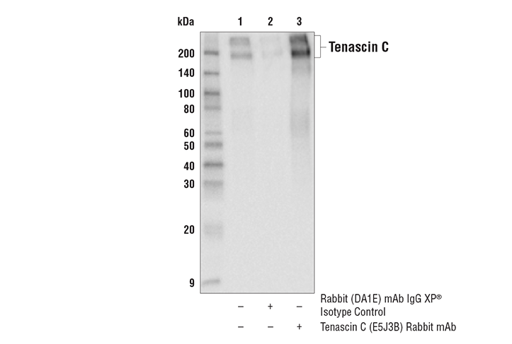
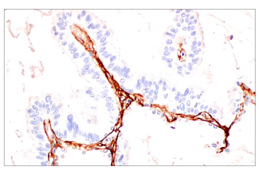
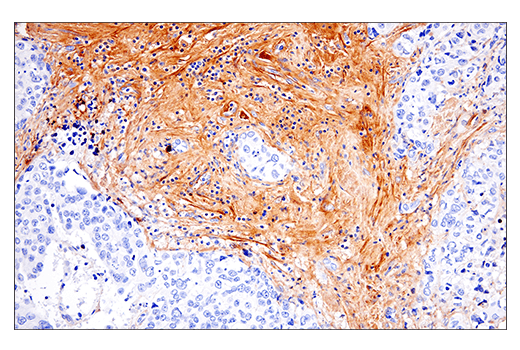
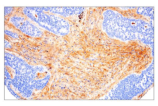
| Tenascin C (E5J3B) Rabbit mAb 33352 | 20 µl |
|
H | 200, 240 | Rabbit IgG |
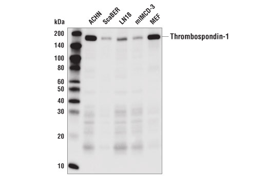
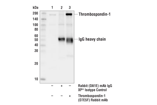
| Thrombospondin-1 (D7E5F) Rabbit mAb 37879 | 20 µl |
|
H M R | 170 | Rabbit IgG |
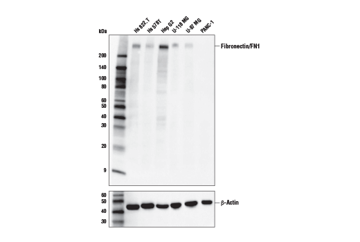
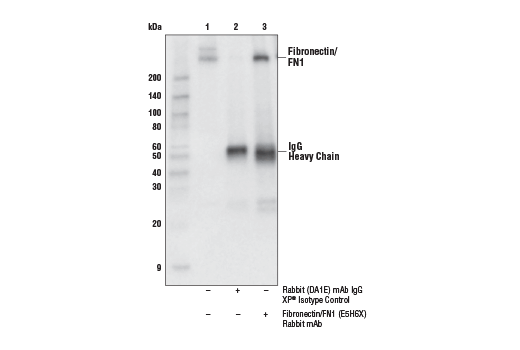
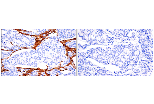
| Fibronectin/FN1 (E5H6X) Rabbit mAb 26836 | 20 µl |
|
H | 300 | Rabbit IgG |
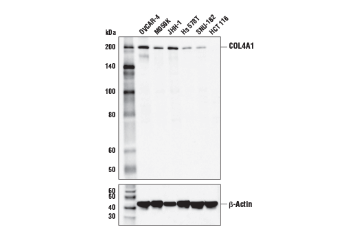
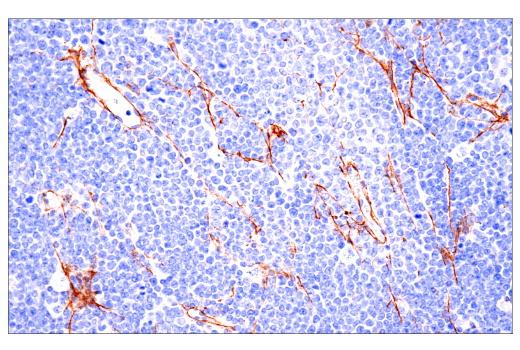
| COL4A1 Antibody 50273 | 20 µl |
|
H | 200 | Rabbit |
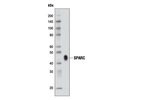
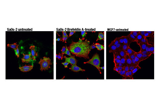
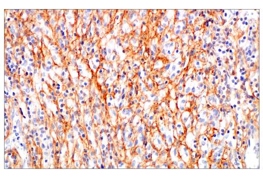
| SPARC (D10F10) Rabbit mAb 8725 | 20 µl |
|
H M | 42 | Rabbit IgG |
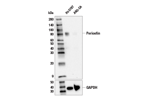
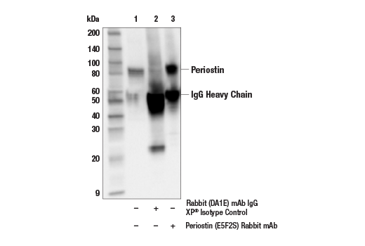
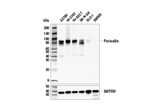
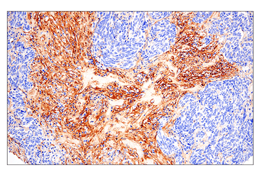
| Periostin (E5F2S) Rabbit mAb 20302 | 20 µl |
|
H | 90 | Rabbit IgG |
| Anti-rabbit IgG, HRP-linked Antibody 7074 | 100 µl |
|
Rab | Goat | |
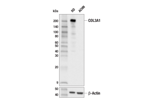
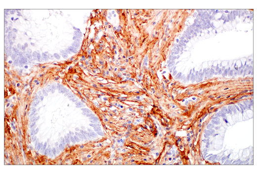
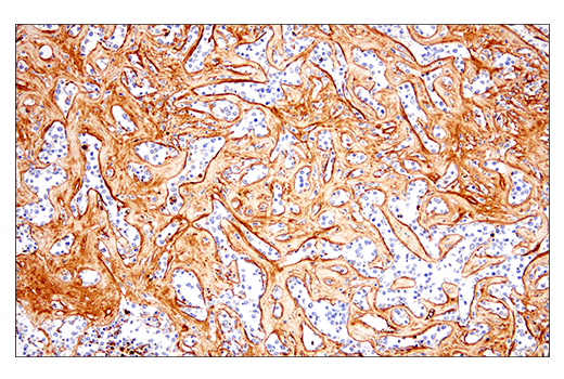
| COL3A1 (E8D7R) XP® Rabbit mAb 63034 | 20 µl |
|
H | 200 | Rabbit IgG |
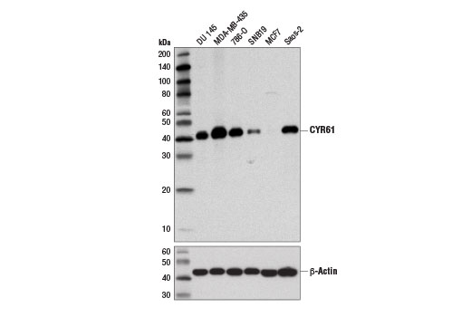
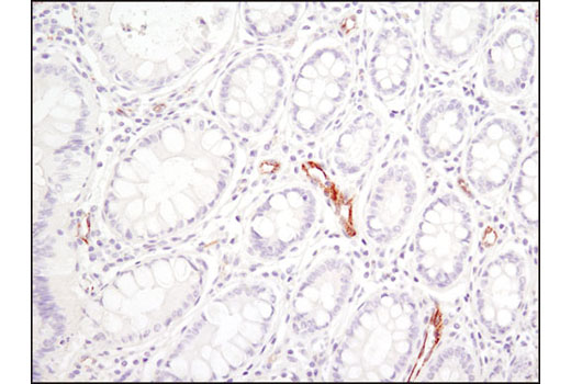
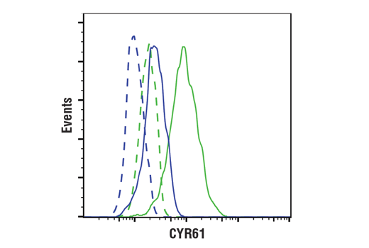
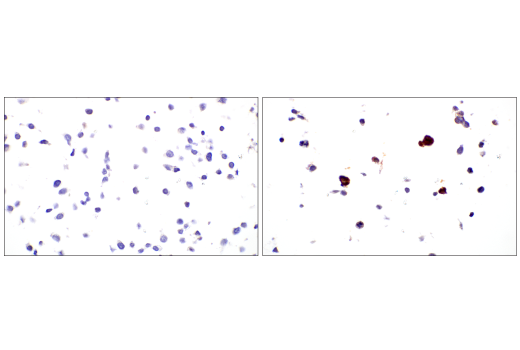
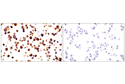
| CYR61 (D4H5D) XP® Rabbit mAb 14479 | 20 µl |
|
H | 41 | Rabbit IgG |
Product Information
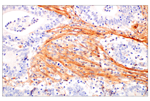
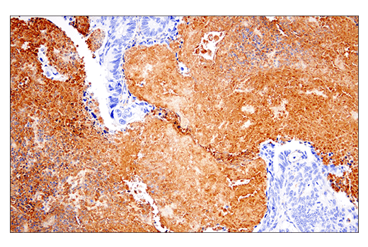
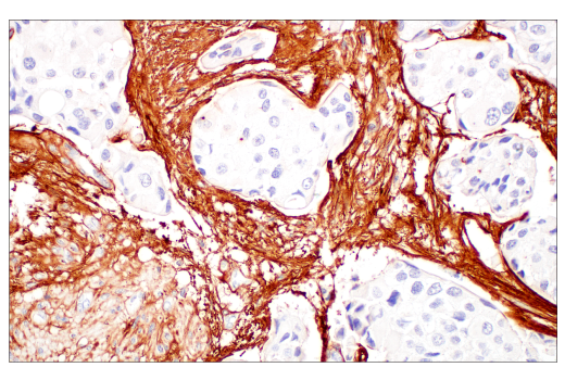
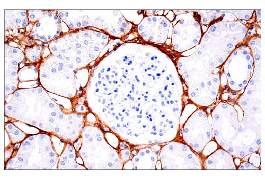
Monoclonal antibodies are produced by immunizing animals with recombinant proteins specific to the carboxy terminus of human Tenascin C protein and human Fibronectin/FN1 protein; with synthetic peptides corresponding to residues surrounding Phe1197 of human COL1A1 protein, Ser395 of human periostin protein, and Pro171 of human CYR61 protein, the amino terminus of human thrombospondin-1 protein and human SPARC protein, and the carboxy terminus of human COL3A1 protein. Polyclonal antibodies are produced by immunizing animals with a synthetic peptide corresponding to residues surrounding Asp536 of human COL4A1 protein. Antibodies are purified by peptide affinity chromatography.
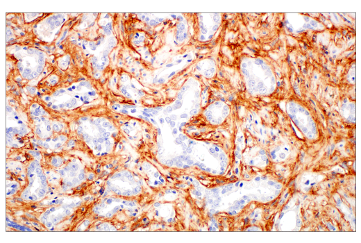
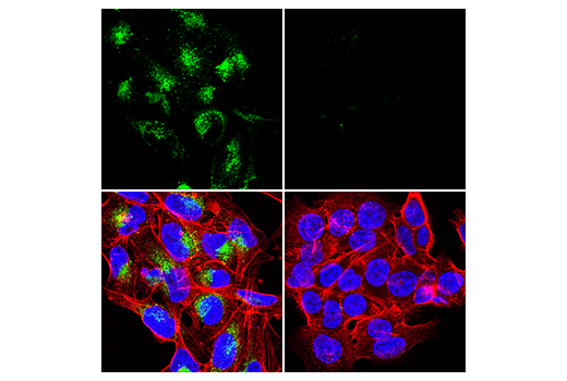
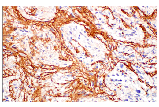
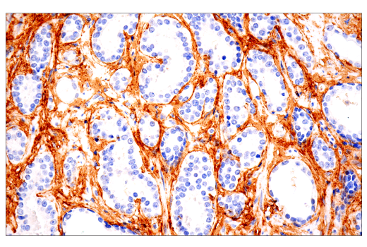
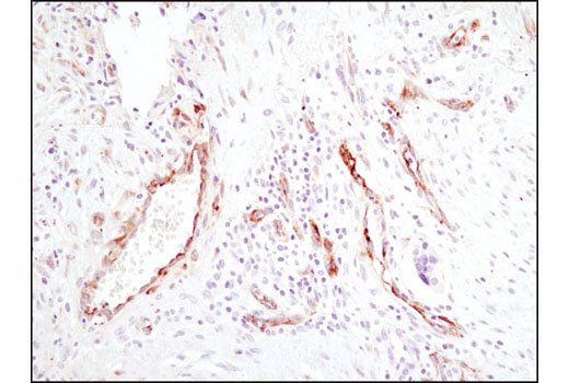
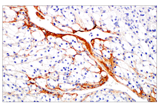
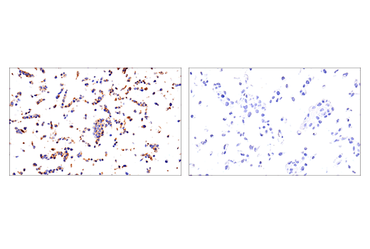
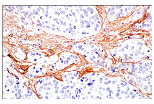
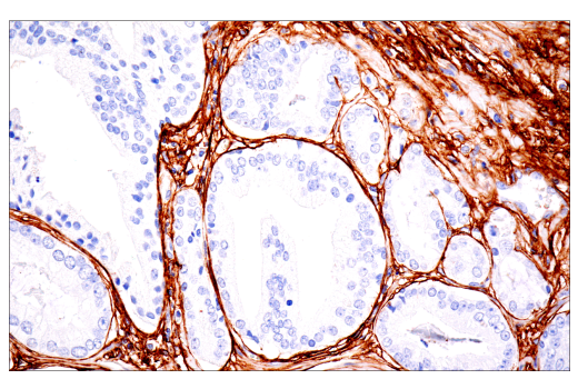
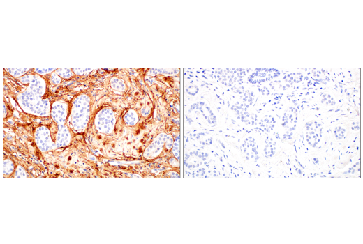
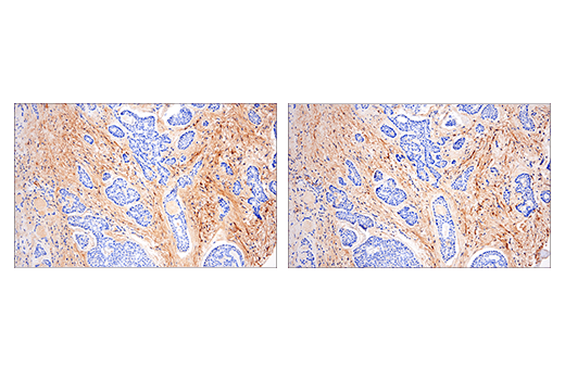
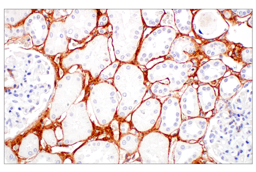
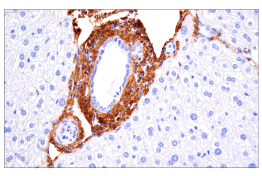
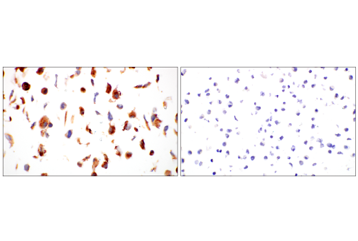
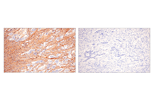
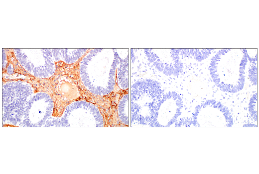
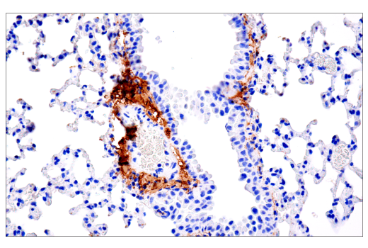
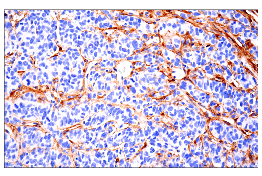
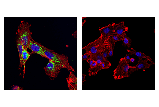
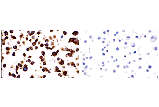
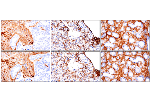
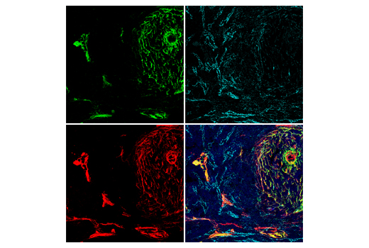
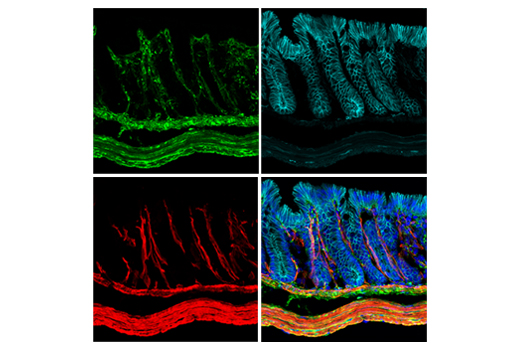
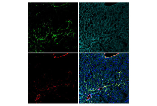
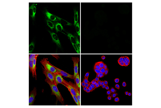
The extracellular matrix (ECM) is a three-dimensional macromolecular network composed of collagens, proteoglycans, glycosaminoglycans, elastin, fibronectin, laminins, along with many other proteins and glycoproteins. This network of macromolecules provides a dynamic microenvironment that supports cell and tissue function, and undergoes continuous remodeling during both normal development and disease (1). Remodeling of the ECM can alter the relative balance of macromolecules within distinct ECM subcompartments, with important functional consequences; for example, changes to the relative amounts of COL1A1 and COL3A1 in the interstitial ECM, or COL4A1 and laminins in the basement membrane, can influence cell-matrix interactions, and/or disrupt cellular signaling events (2). Fibronectin functions as a physical and functional bridge between many different ECM components, including collagens, growth factors, and cell surface integrins, and thus plays a critical role in facilitating ECM remodeling. Matricellular proteins (MCPs) are another important group of ECM proteins. MCPs can be categorized into 6 distinct subgroups: centralized coordination network (CCN), thrombospondin (THBS), secreted protein acidic and rich in cysteine (SPARC), tenascin (TN), small integrin-binding ligand N-linked glycoprotein (SIBLING), and γ-carboxyglutamate (Gla)-containing proteins. CCN1 (CYR61), SPARC, tenascin C, periostin, and thrombospondin-1 are among the most well-studied of this group. All are non-structural ECM proteins that interact with structural ECM proteins, in part to regulate the rigidity of the ECM. They also play important roles in matrix-cell communication by engaging with cell surface receptors and integrins to elicit intracellular responses. The dysregulation of MCP expression has been associated with the development of numerous disease states, including cancer and fibrosis (4,5).
Except as otherwise expressly agreed in a writing signed by a legally authorized representative of CST, the following terms apply to Products provided by CST, its affiliates or its distributors. Any Customer's terms and conditions that are in addition to, or different from, those contained herein, unless separately accepted in writing by a legally authorized representative of CST, are rejected and are of no force or effect.
Products are labeled with For Research Use Only or a similar labeling statement and have not been approved, cleared, or licensed by the FDA or other regulatory foreign or domestic entity, for any purpose. Customer shall not use any Product for any diagnostic or therapeutic purpose, or otherwise in any manner that conflicts with its labeling statement. Products sold or licensed by CST are provided for Customer as the end-user and solely for research and development uses. Any use of Product for diagnostic, prophylactic or therapeutic purposes, or any purchase of Product for resale (alone or as a component) or other commercial purpose, requires a separate license from CST. Customer shall (a) not sell, license, loan, donate or otherwise transfer or make available any Product to any third party, whether alone or in combination with other materials, or use the Products to manufacture any commercial products, (b) not copy, modify, reverse engineer, decompile, disassemble or otherwise attempt to discover the underlying structure or technology of the Products, or use the Products for the purpose of developing any products or services that would compete with CST products or services, (c) not alter or remove from the Products any trademarks, trade names, logos, patent or copyright notices or markings, (d) use the Products solely in accordance with CST Product Terms of Sale and any applicable documentation, and (e) comply with any license, terms of service or similar agreement with respect to any third party products or services used by Customer in connection with the Products.