An orthogonal strategy for antibody validation involves cross-referencing antibody-based results with data obtained using non-antibody-based methods. This approach is critical to verify existing antibody validation data and to identify any effects or artifacts that are directly related to the antibody in question. Providing an additional level of detail to support results generated by the other strategies outlined within this handbook, orthogonal validation often utilizes data that are available in the public domain.
Depending on the antigenic target, non-antibody-based methods can include mining previously published results, studying expression analysis via ‘omics techniques (genomics, transcriptomics, and proteomics), and employing other established antibody-independent methods such as in situ hybridization or RNA sequencing (RNA-seq). Correspondingly, an orthogonal strategy can also be used to ensure that any antibody validation performed inhouse uses the most relevant biological models for the target of interest.
In its simplest form, an orthogonal strategy dictates that results obtained in the other hallmarks require corroboration by non-antibody-based detection methods. As just one example, positive and negative expression of the target observed by binary or ranged strategies should always be confirmed using an orthogonal approach, such as genetic sequencing to confirm knockout or transcriptomic analysis of mRNA to confirm expression
As with all the strategies discussed in this handbook, relying on a single source of information or an individual result is never a good idea. For some targets, there is a significant amount of expression and biological data available (albeit sometimes conflicting) to guide validation strategies. For other targets, it may be necessary to perform appropriate experiments in-house to support antibody testing. Where data from multiple, trusted sources are available, this can, of course, save both time and resources; however, it is important to note that effective orthogonal validation may require additional effort.
Although established immunostaining techniques such as western blot and immunohistochemistry provide a quick visual indication of antibody specificity, it is vital that any antibody validation data generated using these methods are supported by orthogonal testing. One way of achieving this is to mine publicly available databases (eg, CCLE, BioGPS, Human Protein Atlas, DepMap Portal, COSMIC) for genomic and transcriptomic profiling information to help understand whether observed immunostaining results are relevant or are instead due to antibody-related artifacts.
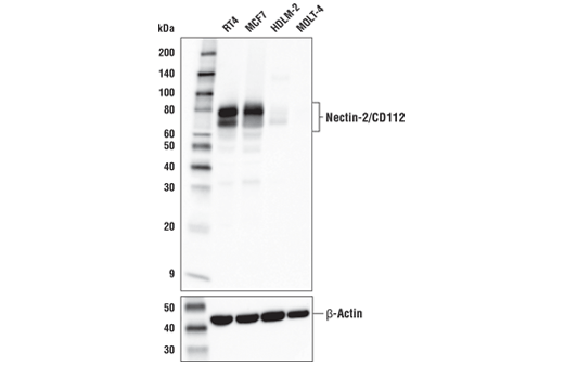
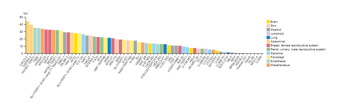
As an example, Figure 1 shows western blot detection of Nectin-2 in a variety of cell lines using the Nectin-2/ CD112 (D8D3F) rabbit mAb. Elevated expression is noticeable in RT4 and MCF7, while only minimal expression can be seen in HDLM-2 and MOLT-4. These results are mirrored by immunohistochemical analysis of RT4 and HDLM-2 cell pellets (Figure 2), clearly illustrating that data from two different applications correlate exceptionally well with the predicted expression of Nectin-2 based on genomics and transcriptomics data, as shown in Figure 3.

Complementing the various ‘omics techniques, orthogonal methods such as in situ hybridization, RNA-seq, and RNAscope allow for the detection of protein expression and/or localization in tissues. These approaches are especially useful for validating antibody data that have been generated using imaging techniques like immunocytochemistry or immunohistochemistry.
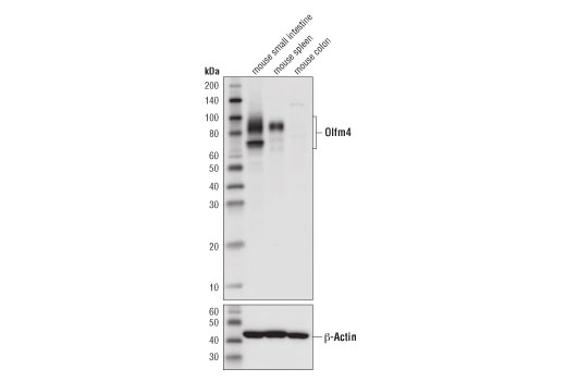
Figure 4 shows IHC analysis of mouse Olfm4 using the Olfm4 (D6Y5A) rabbit mAb. This illustrates positive staining in mouse small intestine and negative staining in colon, a finding which is supported by western blot. (Figure 5). The observed staining pattern is consistent with numerous published orthogonal strategies, including in situ hybridization.1,2
The defining criterion of success for an orthogonal strategy is consistency between the known or predicted biological role and localization of a gene/protein of interest and the resultant antibody staining. This highlights the importance of verifying the specificity and functionality of all reagents in the model and application that will be used in downstream experiments.
Like the other hallmarks described within this handbook, no single validation strategy is sufficient in isolation. Although orthogonal strategies provide evidence that an antibody is behaving as expected, it is critical to combine orthogonal testing with other validation approaches to assure confidence in antibody performance.